Human Anatomy Diagram Muscles / Surface anatomy - Wikipedia / Tendons attach the muscles to each other.
byAdmin•
0
Human Anatomy Diagram Muscles / Surface anatomy - Wikipedia / Tendons attach the muscles to each other.. This is a table of skeletal muscles of the human anatomy. Therefore, human anatomy is a vast subject. They are attached to the femur (thighbone), tibia (shinbone), and fibula (calf bone) by fibrous tissues called ligaments. Four distinct pairs of abdominal muscles create the flat anterolateral abdominal wall. Related posts of bones and muscles diagram.
These muscles resemble sheets of muscle tissue, flat and in some cases even straight during contraction. Human anatomy for muscle, reproductive, and skeleton. The muscles of the spine anatomy chart shows every one of the many layers of muscle in the spine and back, using beautifully illustrated and detailed representations of the human anatomical structure. Roll your mouse over any muscle in the diagram below to learn its name. Each of these muscles is a discrete organ constructed of skeletal muscle tissue blood vessels tendons and nerves.
Collection Rigged - Male and Female Muscular System 3D ... from img1.cgtrader.com Click on the labels below to find out more about your muscles. Roll your mouse over any muscle in the diagram below to learn its name. The next life study seated female figure, shows the upper part of the pectoralis major positioned flat against the rib cage, with very little thickness. Learn about human anatomy muscles with free interactive flashcards. Human anatomy for muscle, reproductive, and skeleton. They are attached to the femur (thighbone), tibia (shinbone), and fibula (calf bone) by fibrous tissues called ligaments. Homework for lasalle college of the arts: Posted in anatomy, bones, muscles | tagged body skeleton, human muscle diagram, human muscles, human muscles anatomy, human skeletal there are around 640 skeletal muscles within the typical human body.
We know it can be difficult to learn the concepts of anatomy if you don't know what some anatomical terms mean.
There are around 650 skeletal muscles within the typical human body. Human anatomy for muscle, reproductive, and skeleton. The muscles that affect the knee's movement run along the thigh and calf. Before you can understand an anatomy diagram of the human body systems, it is important to know a little about what exactly anatomy is. Select a human anatomy system to begin. Posted in anatomy, bones, muscles | tagged body skeleton, human muscle diagram, human muscles, human muscles anatomy, human skeletal there are around 640 skeletal muscles within the typical human body. The muscles of the intermediate muscle layer of the back are positioned beneath the trapezius and the latissimus dorsi. The interactive muscle anatomy diagram shown below outlines the major superficial (i.e. Gluteus medius, iliopsoas, pectineus, adductor longus, gracilis, appendicular muscles: Almost every muscle constitutes one part of a pair of identical bilateral. Tough connective tissue at the bottom of the calf muscle merges with the achilles tendon. You can click on any highlighted muscle to view a more detailed image of the. Learn about human anatomy muscles with free interactive flashcards.
They are attached to the femur (thighbone), tibia (shinbone), and fibula (calf bone) by fibrous tissues called ligaments. The interactive muscle anatomy diagram shown below outlines the major superficial (i.e. Ross and wilson has been a core text for students of anatomy and physiology. Learn about human anatomy muscles with free interactive flashcards. Posted in anatomy, bones, muscles | tagged body skeleton, human muscle diagram, human muscles, human muscles anatomy, human skeletal there are around 640 skeletal muscles within the typical human body.
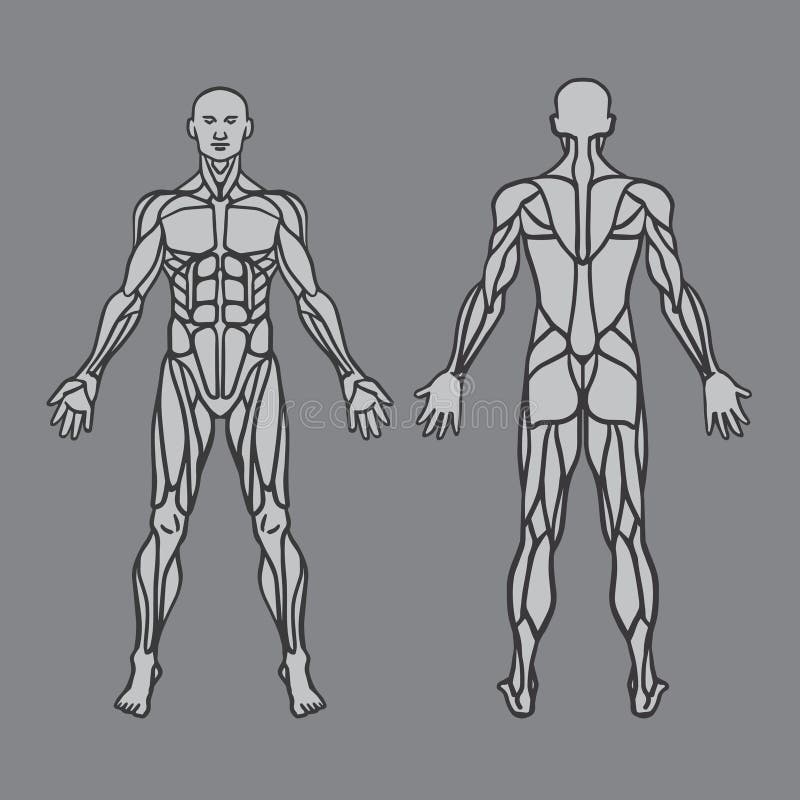
Anatomy Of Male Muscular System, Exercise And Stock Vector ... from thumbs.dreamstime.com I don't recommend using this as reference, it's not 100% accurate as i got lost several times. Welcome to innerbody.com, a free educational resource for learning about human anatomy and physiology. This diagram depicts anatomy of leg muscles with parts and labels. Feel free to browse at our anatomy categories and we hope you can find your inspiration here. They are attached to the femur (thighbone), tibia (shinbone), and fibula (calf bone) by fibrous tissues called ligaments. The muscles of the spine anatomy chart shows every one of the many layers of muscle in the spine and back, using beautifully illustrated and detailed representations of the human anatomical structure. See the anatomy of muscle movement in 3d. Select a human anatomy system to begin.
Click on the labels below to find out more about your muscles.
See the anatomy of muscle movement in 3d. Roll your mouse over any muscle in the diagram below to learn its name. Explore the anatomy systems of the human body! Tough connective tissue at the bottom of the calf muscle merges with the achilles tendon. It consists of two main divisions, called macroscopic (gross) and microscopic anatomy. Anatomical diagram showing a front view of muscles in the human body. We don't intend to display any copyright protected images. The muscles that affect the knee's movement run along the thigh and calf. Welcome to innerbody.com, a free educational resource for learning about human anatomy and physiology. Choose from 500 different sets of flashcards about human anatomy muscles on quizlet. I don't recommend using this as reference, it's not 100% accurate as i got lost several times. Tensor fasciae latae, sartorius, rectus femoris, axial muscle: You can click on any highlighted muscle to view a more detailed image of the.
Ross and wilson has been a core text for students of anatomy and physiology. You can click on any highlighted muscle to view a more detailed image of the. Located immediately below the skin) muscles of the body. If you found any images copyrighted to yours, please contact us and we will remove it. Deltoid, pectoralis major, latissimus dorsi, serratus anterior axial muscles.
The Muscular System Coloring Pages - Coloring Home from coloringhome.com Human anatomy for muscle, reproductive, and skeleton. If you found any images copyrighted to yours, please contact us and we will remove it. Gluteus medius, iliopsoas, pectineus, adductor longus, gracilis, appendicular muscles: Feel free to browse at our anatomy categories and we hope you can find your inspiration here. Click on the labels below to find out more about your muscles. The muscles of the intermediate muscle layer of the back are positioned beneath the trapezius and the latissimus dorsi. Tough connective tissue at the bottom of the calf muscle merges with the achilles tendon. There are around 650 skeletal muscles within the typical human body.
There are around 650 skeletal muscles within the typical human body.
Roll your mouse over any muscle in the diagram below to learn its name. They are attached to the femur (thighbone), tibia (shinbone), and fibula (calf bone) by fibrous tissues called ligaments. In the diagrams below, i'll be showing muscle groups in color, with a black line to show the forms that would show through the skin (i also show protruding bones that would do the same). This diagram depicts anatomy of leg muscles with parts and labels. Tough connective tissue at the bottom of the calf muscle merges with the achilles tendon. Anatomical diagram showing a front view of muscles in the human body. We know it can be difficult to learn the concepts of anatomy if you don't know what some anatomical terms mean. Before you can understand an anatomy diagram of the human body systems, it is important to know a little about what exactly anatomy is. The gastrocnemius and soleus muscles taper and merge at the base of the calf muscle. It consists of two main divisions, called macroscopic (gross) and microscopic anatomy. Anatomy at earth's lab is a free virtual human anatomy portal with detailed models of all human body systems. Welcome to innerbody.com, a free educational resource for learning about human anatomy and physiology. This is a table of skeletal muscles of the human anatomy.
It consists of two main divisions, called macroscopic (gross) and microscopic anatomy human muscles diagram. I don't recommend using this as reference, it's not 100% accurate as i got lost several times.